What is your area of research specialism and what attracted you to it?
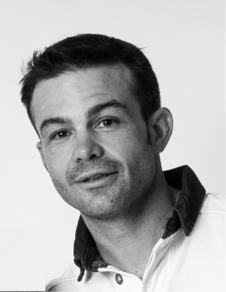
I’m working on magnetic resonance imaging (MRI), mainly diffusion MRI applied to cerebral development. Diffusion imaging is a unique technique to probe and characterise the brain microstructure non-invasively. After a master’s degree in physics, I turned to medical imaging during my PhD because I wanted to do science oriented towards human beings. After my PhD in France, I joined Professors Petra Hüppi and Stéphane Sizonenko in Geneva to work on development and application of diffusion imaging to perinatal brain injuries. Cerebral development is an incredibly interesting process and to be able to get insight the brain with MRI is even more interesting.
What has been your proudest research accomplishment so far?
Alongside international collaborators, we were able to develop and characterise a sheep model of periventricular leukomalacia, a type of brain injury that affects premature infants. Being gyrified, the sheep brain is closer to the human brain and, as such, lesions observed on animal models on the sheep are also closer to the lesions seen on human babies. This is very important for understanding injury mechanisms as well as testing neuroprotective strategies. MRI is an essential tool in this context.
What is the most important question you want to address in your research?
The development of diffusion imaging techniques to best model biophysical changes in the brain is one of my research topics of high interest. But all this pre-clinical research has only one goal: to improve the outcome and quality of life of babies born prematurely.
What is your lab’s role on the PREMSTEM project?
My institution, Université de Genève, is part of the centre of biomedical imaging in Lausanne (Switzerland), a laboratory where we have state-of-the-art MRI scanners with two small bore magnets at ultra-high magnetic field (9.4T and 14.1T versus 3.0T used routinely in clinics). We have huge experience in MRI and spectroscopy. As such, all of the MRI on rodents used in the PREMSTEM project will be done in our lab. Furthermore, we will bring our expertise to MRI scans performed on larger animals (i.e. sheep) to help implement advanced imaging techniques.
What is innovative about PREMSTEM? Why is this research important?
This research is important because the aim is to save or improve the quality of life of babies born prematurely. The idea is to give tools to clinicians to heal these children and consequently improve their lives as well as the lives of their loved ones. The impact of this research is actually huge.
What is the most significant outcome you hope PREMSTEM can achieve?
To find a treatment that can be used in neonatology to ensure normal development in preterm babies. The consequences for society will be multiple: lower hospital costs (due to a shorter length of stay), less long-term care, less suffering for families… and healthy babies!


