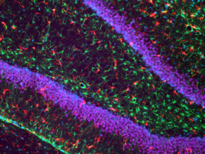
Magnification of the rat brain at the level of the hippocampus
Microglia in red, astrocytes in green, and neurons in purple with blue used as a counterstain.
Credit: Chantal Kosmeijer, UMC Utrecht
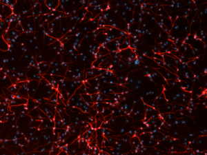
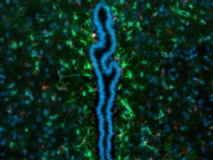
Neural stem cells differentiated in vitro towards neurons
Neural stem cells differentiated in vitro towards neurons which form networks in a dish, with blue used as a counterstain.
Credit: Sara De Palma, UMC Utrecht
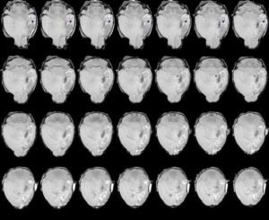
Mouse brain with hypoxic-ischemic brain injury
A montage created using MRI showing hypoxic-ischemic brain injury.
Credit: Sara De Palma and group of Rick Dijkhuizen (Annette van der Toorn and Geralda van Tilborg), UMC Utrecht
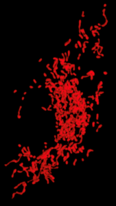
Hippocampal neurons after hyperoxia
Hippocampal neurons after hyperoxia, a state where there is too much oxygen, cultured together with human mesenchymal stem cells (h-MSCs).
Credit: Meray Sendar, Universitätsmedizin Essen
Mitochondria inside a single microglia cell
Credit: Syam Nair, University of Gothenburg
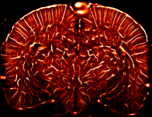
Vascular density of a complete coronal section of the living rat brain
Credit: Iconeus
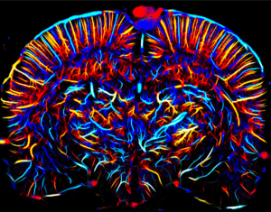
Axial velocities of gas microbubbles
Measured in the mm/s range and their direction (blue is upward flow, red is downward flow).
Credit: Iconeus
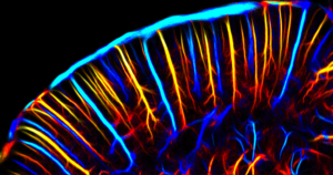
Close-up of the cortex
This parameter allows for discrimination between arterial flow that perfuse the tissues (in red) and venous outflow (in blue).
Credit: Iconeus
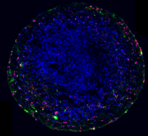
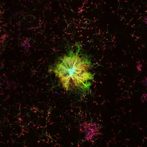
Cortical brain organoid (mini brain)
Organoid cultured from human induced pluripotent stem cells (derived from skin biopsy samples) showing immature (red) and mature (red+green) oligodendrocytes. Mature oligodendrocytes will start to produce myelin in the mini-brain. All cell nuclei are stained in blue.
Credit: Myrna Brandt and Sebastiaan Corstjens, UMC Utrecht
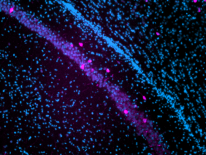
Interneurons in the hippocampus
Parvalbumin-positive interneurons (pink) in the CA2 area of the hippocampus in the rat brain. All cell nuclei are counterstained with DAPI (blue).
Credit: Judit Alhama Riba, UMC Utrecht
Microglial cells and astrocytes
Microglial cells (red) and astrocytes (green) located at the third ventricle in the rat brain. All cell nuclei are counterstained with DAPI (blue).
Credit: Judit Alhama Riba, UMC Utrecht
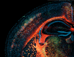
Cortex and hippocampus of the rat brain
Stained in red with N-acetylgalactosamine-binding Wisteria floribunda agglutinin to visualise perineural nets (myelin specifically lights up) and in green for parvalbumin-positive interneurons. Counterstain of all cell nuclei with DAPI in blue.
Credit: Chantal Kosmeijer, UMC Utrecht